Tomocube
Label-free quantitative imaging:
An innovative approach to exploring cells and tissues
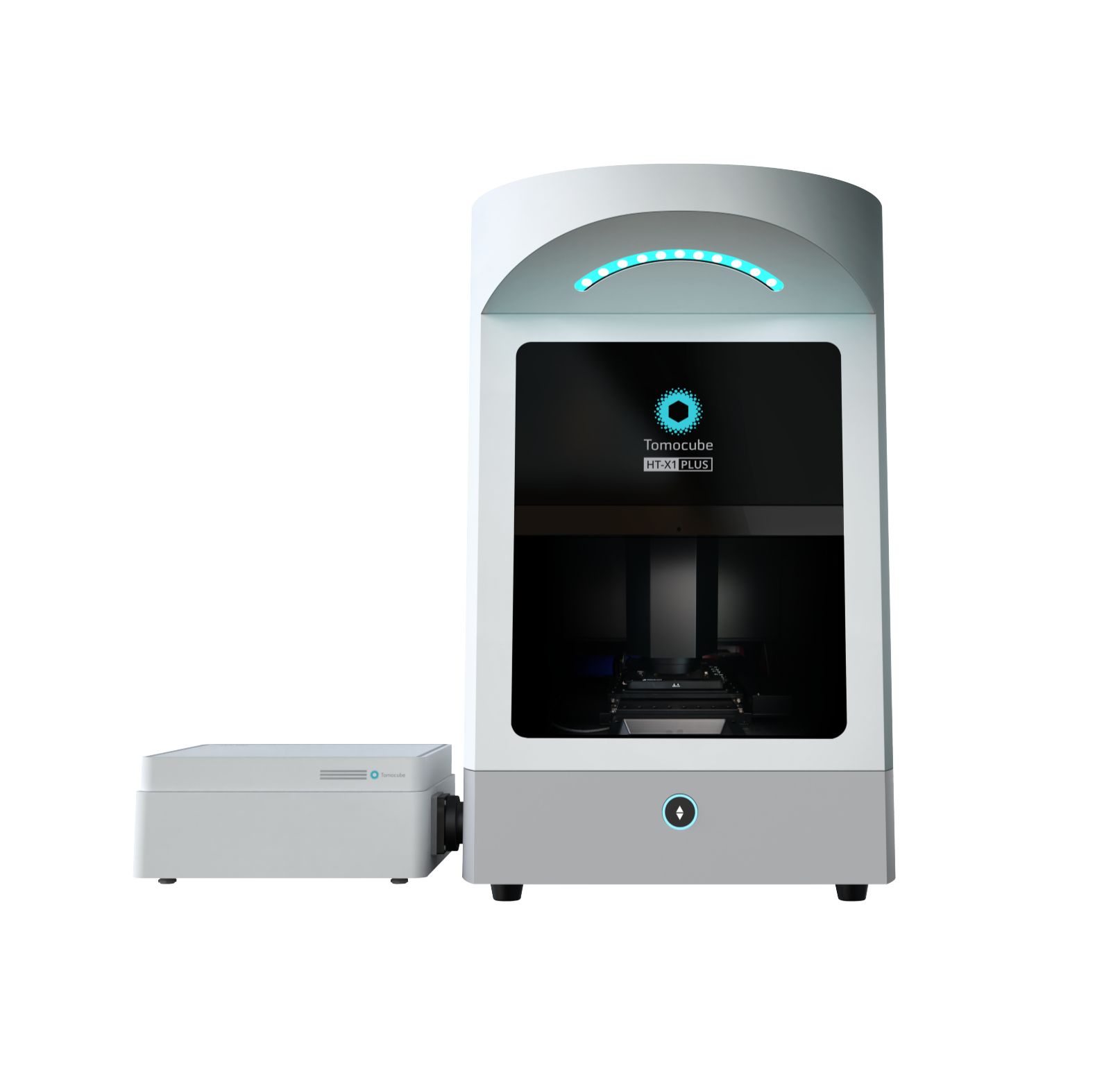
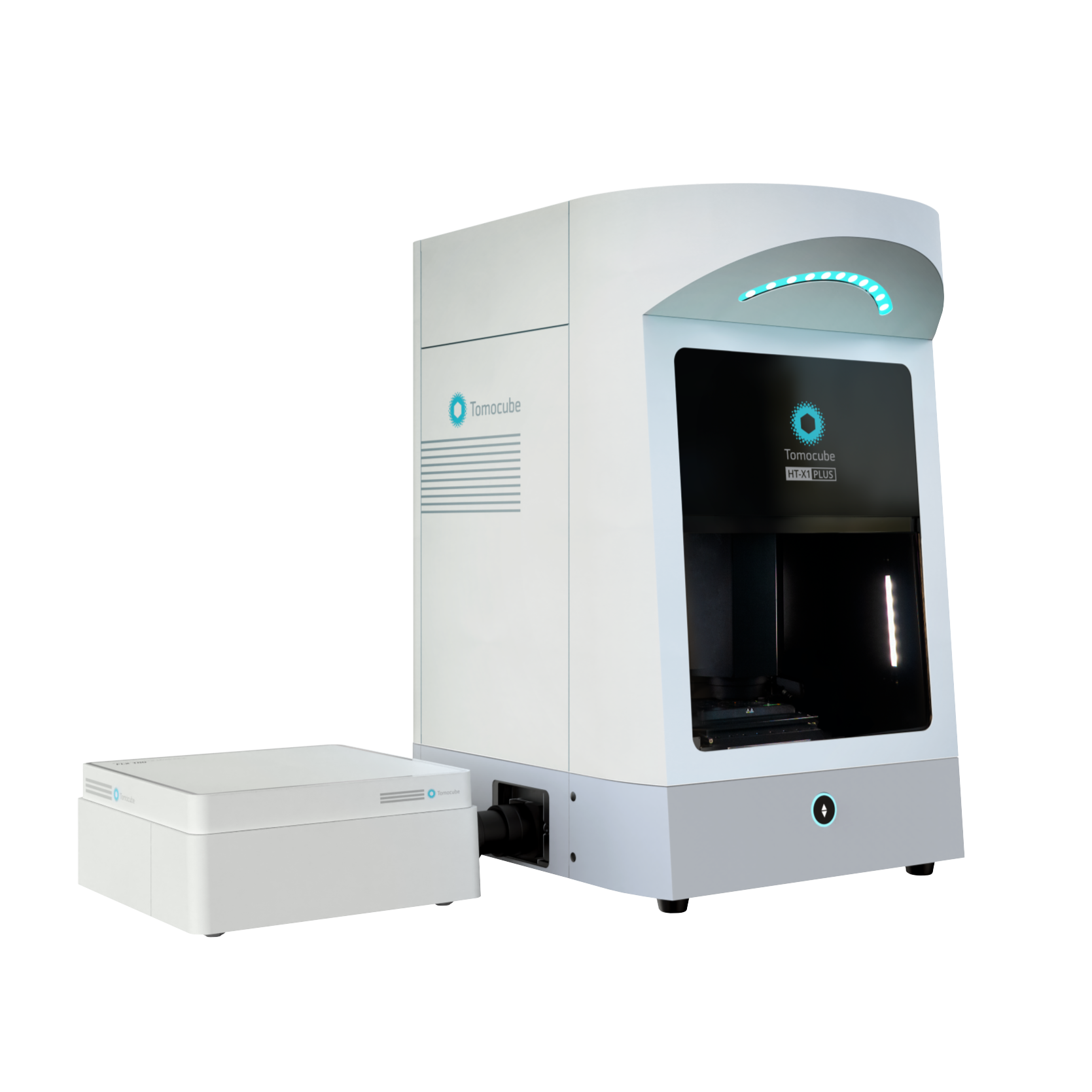
Holotomography(HT)
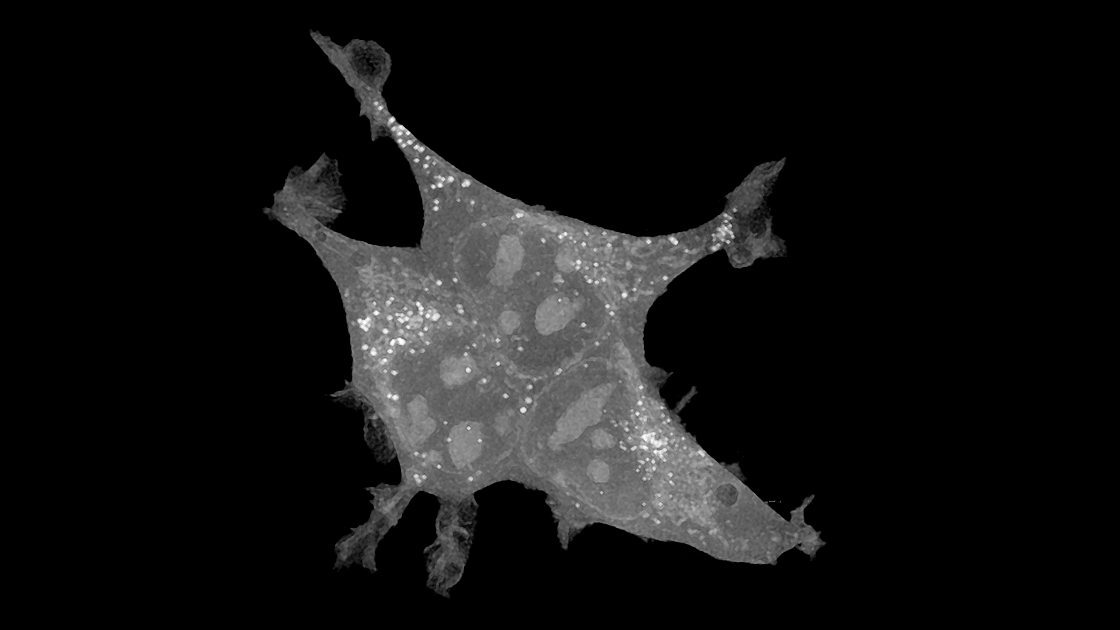
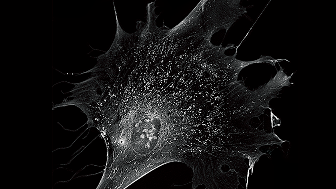
HT illuminates a cell with very low power visible light at various illumination angles and measures the phase delay of the transmitted light. Although optically similar to CT, it differs in that it uses measured refractive index (RI) as imaging contrast, allowing visualization of living cells and tissues without labeling or staining.
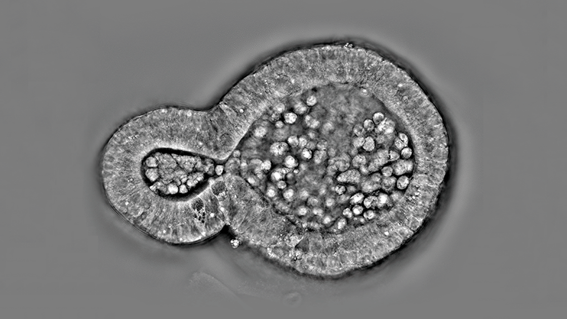
Just as a CT scan uses X-ray absorptivity as the imaging contrast to see inside a patient’s organs without invasive procedures, HT uses the refractive index (RI), an intrinsic optical parameter describing the speed of light passing through a specific material, to visualize living cells and tissues. Since variations in the biomolecular concentration directly impact the overall RI of the cellular biomaterials, the reconstruction of RI tomogram by Holotomography can retrieve important biophysical parameters such as cell volume, surface area, or protein concentration for further analysis.
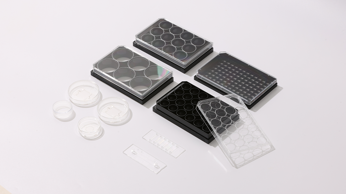
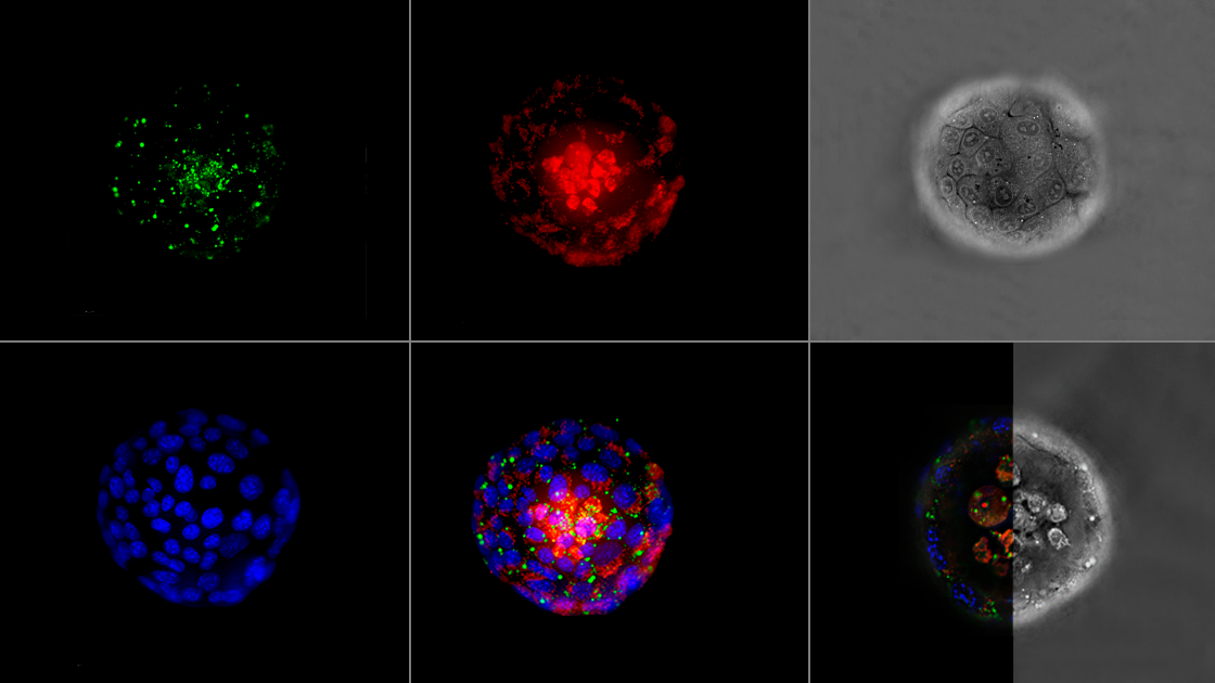
Holotomography’s applications span from cellular components like nuclei and mitochondria to studying organoids, tissues, and even whole organisms, enabling breakthroughs in cellular biology understanding. Its applications continue to expand as new advances are made in tomographic imaging.