 Image 1 of 5
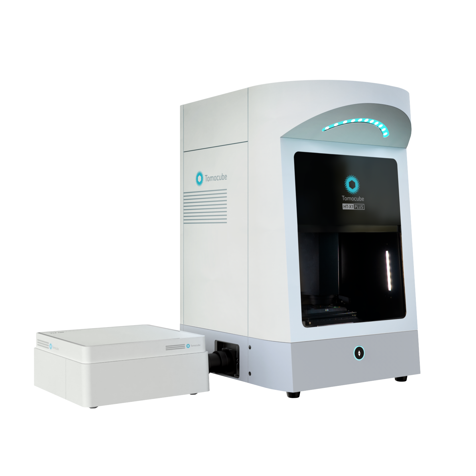 Image 2 of 5
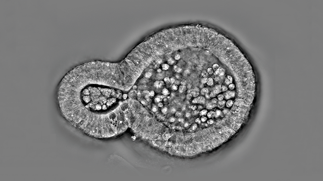 Image 3 of 5
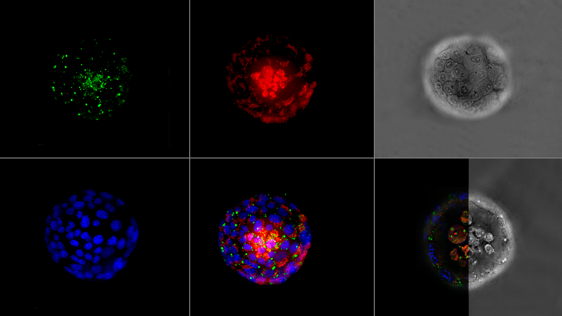 Image 4 of 5
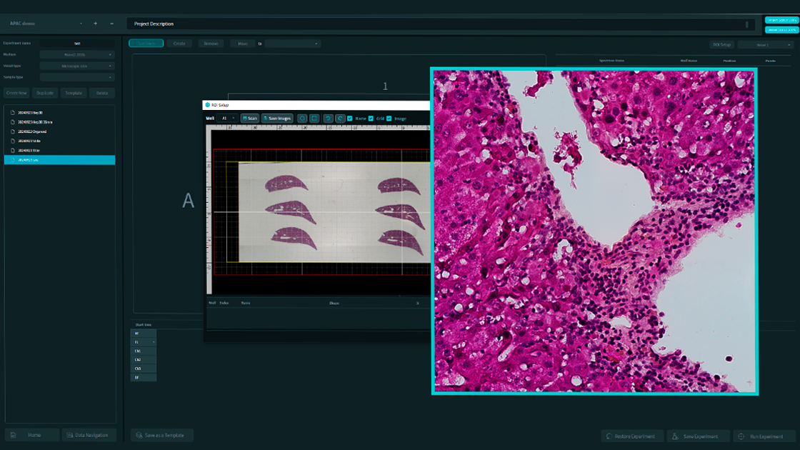 Image 5 of 5
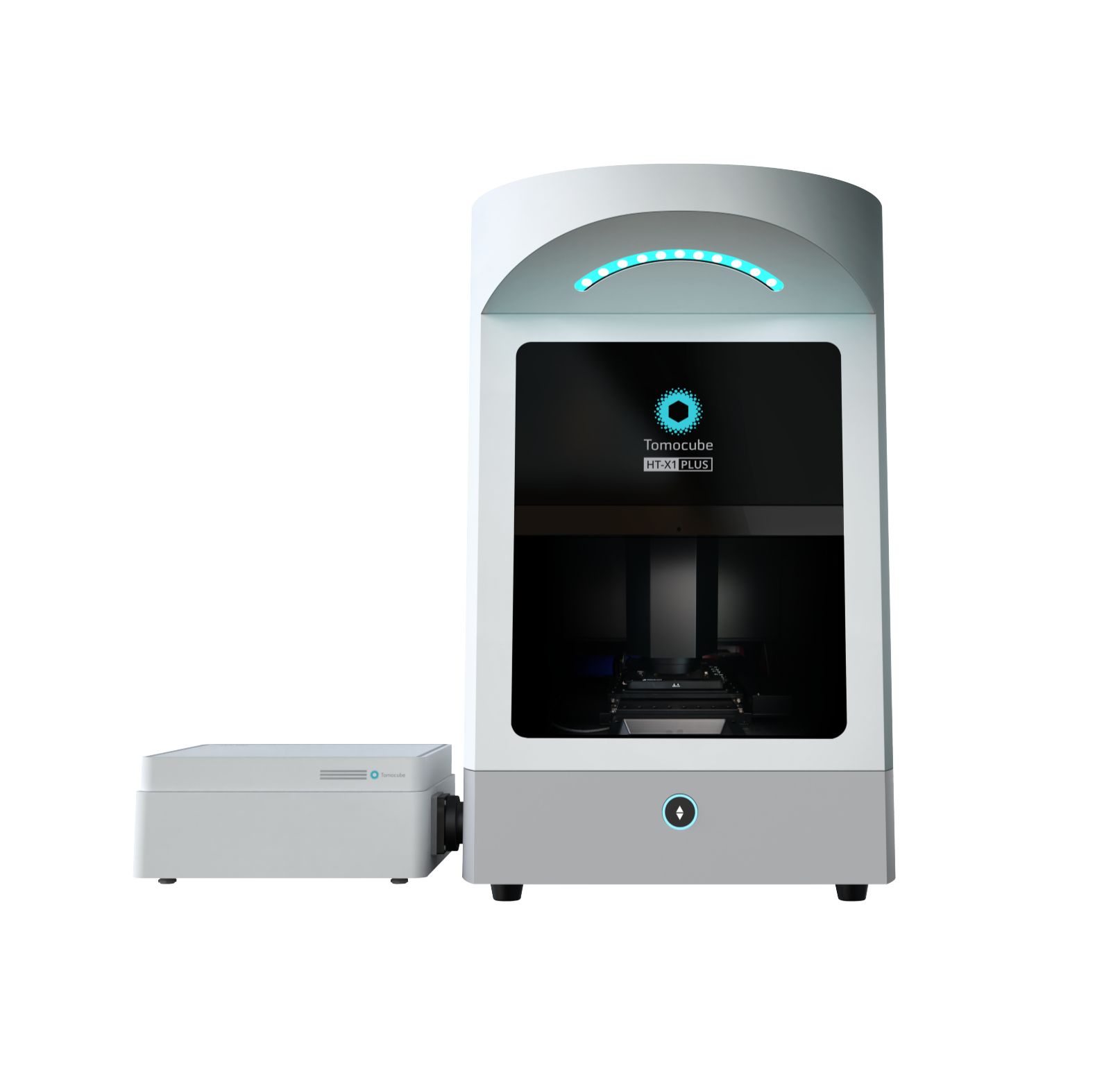
The HT-X1™ Plus takes bioimaging to the next level, building on the proven success of the HT-X1™.
As the first of its kind in the 2nd-generation Holotomography series, the HT-X1™ revolutionized biomedical research with its high-resolution, 3D imaging and unparalleled stability. Its versatile platform, compatible with various imaging plates and powered by the advanced TomoAnalysis™ software, provided researchers with powerful tools for detailed quantitative analysis and reliable imaging across a wide range of applications.
Equipped with a high-spec camera featuring a 4x larger field of view and significantly reduced acquisition time, the HT-X1™ Plus is perfect for high-throughput phenotypic screening of cells and organoids. Its upgraded correlative imaging capabilities—incorporating an sCMOS-based fluorescence module—enable seamless integration of molecular studies with single-cell-resolution 3D images.
The HT-X1™ Plus extends the reach of Holotomography to an even broader array of challenging specimens, including dense organoids, tissue sections, and fast-moving microorganisms. It is a state-of-the-art Holotomography imaging platform, designed to empower researchers with the precision, efficiency, and reliability needed to drive the future of biological and biomedical discovery.
The HT-X1™ Plus is optimized for high-throughput screening, making it highly suitable for high-content, image-based drug screening research. Featuring a high-performance CXP camera and AI-powered image reconstruction algorithms, the platform excels in both coverage and acquisition speed. Its large field of view measuring 308 μm x 308 μm and rapid 3D scanning capability allows researchers to efficiently analyze an entire 96-well plate in under 30 minutes. This efficiency enables large-scale experiments with unmatched precision and consistency, resulting in faster, more reliable data acquisition that significantly accelerates the drug discovery process.
With the new color brightfield imaging modality and wide preview scan features, researchers can gain deeper insights into tissue section studies. The platform allows for the seamless integration of complex structural data, obtained through 3D optical sectioning, with rich histological information from H&E staining or immunohistochemistry. This integration enhances our understanding of tissue morphology and dynamics, accelerates advancements in clinical pathology and diagnostics, and helps pave the way for the future of personalized medicine.
The HT-X1™ Plus offers enhanced multimodal imaging capability with its fluorescence module (FLX™) featuring an sCMOS camera designed specifically for precise signal intensity measurements. The FLX™ module offers high sensitivity to fluorescence, achieving better signal-to-noise ratio (SNR) and shorter exposure times. This allows researchers to obtain biomolecular specificity information from target organelles or fluorescence sensors, even in samples with weak fluorescent signals, such as antibody reactions or hard-to-stain organoids.
Features:
Large Field-of-view
Faster Image Acquisition
Flexible Choice of Light Source
Combine Advanced Fluorescence
Wide Preview + Color Brightfield