 Image 1 of 4
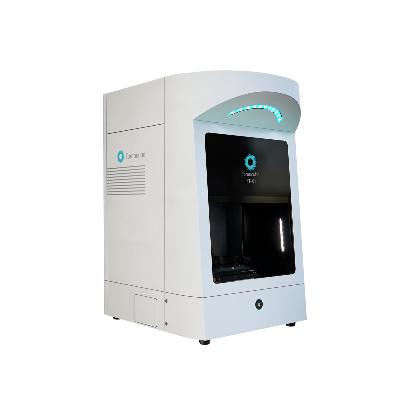 Image 2 of 4
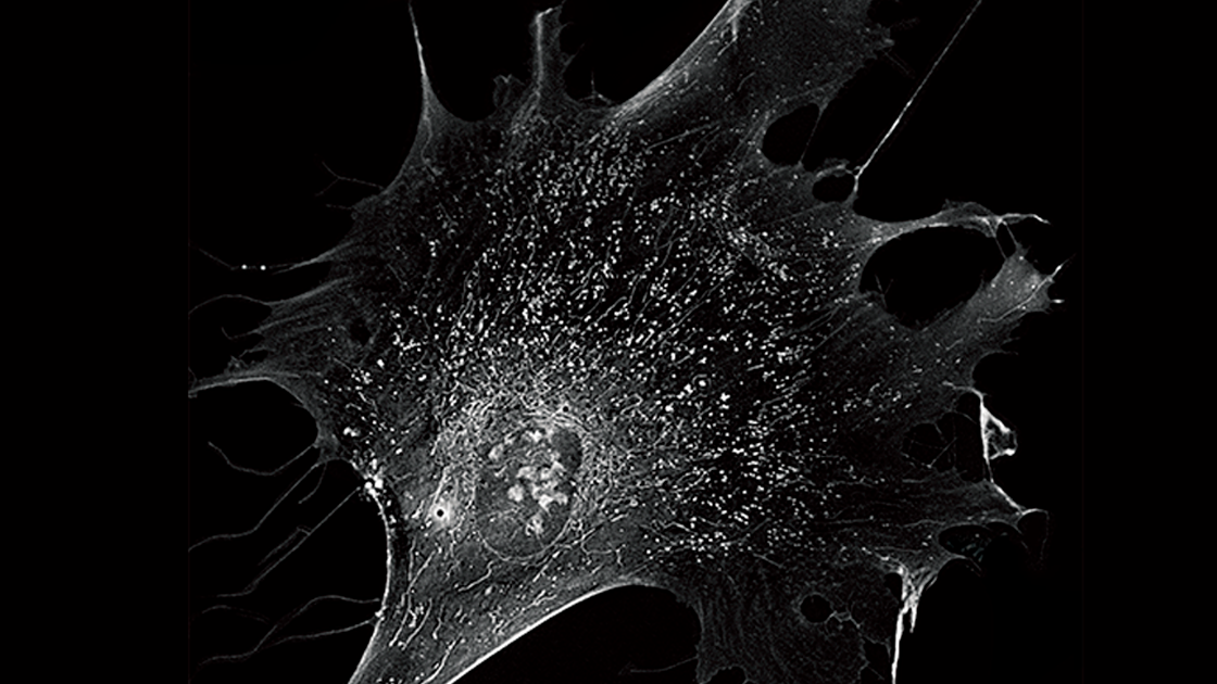 Image 3 of 4
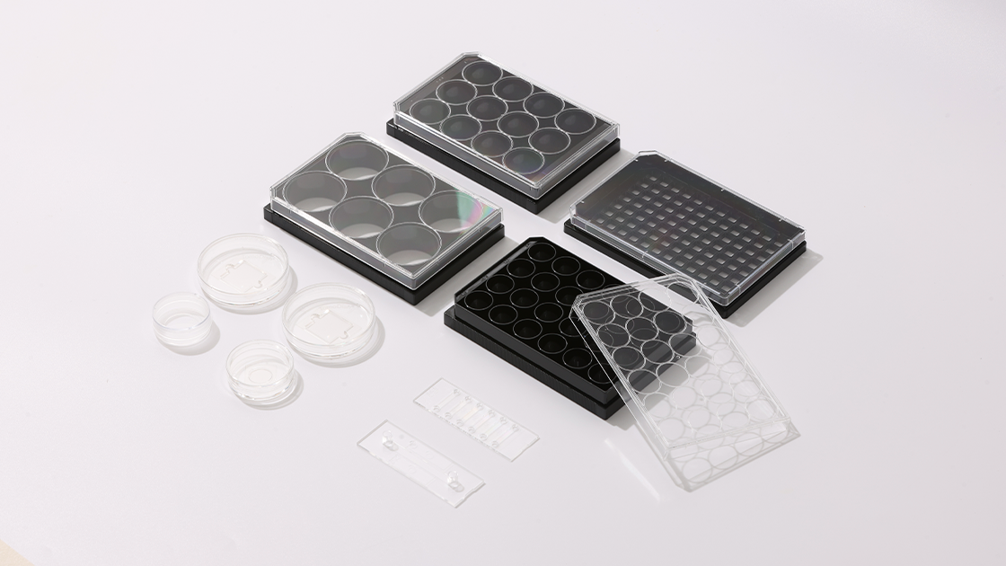 Image 4 of 4
The #1 Choice for Live-Cell Imaging and Analysis
Empowering your Exploration. Redefining the Limits.
Holotomography is a technique that utilizes low-intensity light to acquire the refractive index of cells from multiple angles. This technology has emerged as an excellent cellular imaging solution, allowing for high-resolution imaging while maintaining the health of the cells by using low-intensity light sources. Tomocube's second-generation Holotomography, the Tomocube HT-X1, employs low-coherence light sources, ensuring lower toxicity and noise-free high-resolution image acquisition compared to laser-based methods.
The high-resolution images obtained through this technology enable real-time observation of not only the morphology of living cells but also the shapes of subcellular organelles, such as the nucleus, nucleolus, mitochondria, and lipid droplets.
The integrated stage-top incubator provides a stable cultivation environment, enabling prolonged monitoring of sensitive cells like stem cells or organoids.
Especially designed to maximize user flexibility, the HT-X1 employs a unique adaptive illumination module that is tailored for multi-well plates. The combination of high NA, a long working distance condenser, DMD, and a motorized illumination unit delivers an efficient illumination pattern for a diverse range of vessel types, from 35-mm dishes to 96-well plates. With the HT-X1, researchers will have more freedom to design their experiments on different sample types using any imaging vessel of their choice.
TomoStudio X works in concert with the HT-X1 platform to visualize and analyze RI tomograms. This intuitive, easy-to-use software empowers users with complete system control, allowing them to handle even the most complex experiments through simple mouse clicks. Researchers can easily set up complex time-lapse sequences, perform large-area tile imaging and stitching tasks, and image multiple points with ease using this software.
Features:
Label-free 3D visualization
Correlative fluorescence
Long-term timelapse
High-throughput screening
Quantitative analysis